Long Bone Labeled Endosteum - Periosteum Anatomy Britannica - See bone and cartilage development.. Among these cells, you can find the bone stem cells, the ones that are going to further develop into osteoblasts and osteoclasts. The endosteum (plural endostea) is a thin vascular membrane of connective tissue that lines the inner surface of the bony tissue that forms the medullary cavity of long bones. The first ones are cells that contribute to the formation of bone, while the latter represent. Osteoclasts on the inside in the endosteum remove this bone to maintain the bone diameter. Transcribed image text from this question.
Label the structures of a long bone medullary epiphyseal cavity line spongy articular bone cartilage periosteum compact bone endosteum. The endosteum (plural endostea) is a thin layer of connective tissue which lines the surface of the bony tissue that forms the medullary cavity of long bones. Lesson #39 presented long bone anatomy, but let's take a moment to review. Bone and cartilage at rosalind franklin university these pictures of this page are about:long bone endosteum. Observe regions of trabecular bone and cortical bone in this specimen.
Schematic Diagram Of Long Bone Cross Section 47 Download Scientific Diagram from www.researchgate.net The endosteum is in the marrow cavity. Osteoclasts of the endosteum remove bone from the inside so the thickness remains fairly constant, a highly regulated process. Terms in this set (12). Furthermore, on histological sections, fluorescently labeled lin−sca1+kit+ hspc from. Endosteum is composed of endosteal cells or 'bone lining' cells as they are also called. Long bone endosteum (page 1). These are strong bones because they must be able to withstand the force generated when endosteum lines the inner surface of the medullary cavity of all long bones. Structure of long bone although there are many different types of bones in the skeleton, we will discuss the different parts of a optional activity:
The outer surface of compact bone is covered with a fibrous material called periosteum to which muscles attach.
Bone and cartilage at rosalind franklin university these pictures of this page are about:long bone endosteum. The endosteum (plural endostea) is a thin layer of connective tissue which lines the surface of the bony tissue that forms the medullary cavity of long bones. The first ones are cells that contribute to the formation of bone, while the latter represent. Long bones — a subtype of bones — are longer than they are wide. The outer surface of compact bone is covered with a fibrous material called periosteum to which muscles attach. Draw and label a longitudinal section of a long bone. Label the structures of a long bone medullary epiphyseal cavity line spongy articular bone cartilage periosteum compact bone endosteum. This endosteal surface is usually resorbed during long periods of malnutrition, resulting in less cortical thickness. Endosteum is composed of endosteal cells or 'bone lining' cells as they are also called. Lesson #39 presented long bone anatomy, but let's take a moment to review. Among these cells, you can find the bone stem cells, the ones that are going to further develop into osteoblasts and osteoclasts. Terms in this set (12). The endosteum (plural endostea) is a thin vascular membrane of connective tissue that lines the inner surface of the bony tissue that forms the medullary cavity of long bones.
Image h shows in detail the distribution of bone cells in. Osteoclasts of the endosteum remove bone from the inside so the thickness remains fairly constant, a highly regulated process. The diaphysis is the hollow, tubular shaft that runs between interior of each long tubular bone of the limbs presents a cylindrical cavity named marrow cavity and it is lined with the medullary membrane called endosteum. This video was produced to help students of human anatomy at modesto junior college study our anatomical models. Draw and label a longitudinal section of a long bone.
Long Bone Labeled Endosteum Adult Long Bone Sagittal Section Through Long Bone Showing The Download Scientific Diagram Membranes Including The Endosteum And Periosteum from i0.wp.com If medullary lesions develop along the inner aspect of the cortical bones, especially in the long bones. The end of the long bone is the epiphysis and the shaft is the diaphysis. This image represents the parts of a long bone. Long, short, flat, irregular and sesamoid. The endosteum (plural endostea) is a thin layer of connective tissue which lines the surface of the bony tissue that forms the medullary cavity of long bones. The endosteum is also medically termed as the medullary membrane, located in the diaphysis (cavity of long bones). Initially, multiple epitheloid cell granulomas or granulomatous lesions containing fibrin deposits began to appear in the. Bone and cartilage at rosalind franklin university these pictures of this page are about:long bone endosteum.
Long bone endosteum (page 1).
Long bones are those that are longer than they are wide. The diaphysis is the hollow, tubular shaft that runs between interior of each long tubular bone of the limbs presents a cylindrical cavity named marrow cavity and it is lined with the medullary membrane called endosteum. Bone and cartilage at rosalind franklin university these pictures of this page are about:long bone endosteum. Furthermore, on histological sections, fluorescently labeled lin−sca1+kit+ hspc from. When osteoclasts start removing less bone, or osteoblasts start adding more bone, the. The inner surface is called endosteum. The inner circumferential lamella is labeled. Structure of long bone although there are many different types of bones in the skeleton, we will discuss the different parts of a optional activity: It is important to note that the absence of endosteum or periosteum on a bone signals that the bone is ready to be reabsorbed by correct answer 2. The delicate connective tissue layer lining the inside surface of compact bone. The first ones are cells that contribute to the formation of bone, while the latter represent. Image h shows in detail the distribution of bone cells in. Terms in this set (12).
Definition and functions the endosteum is a structure in the middle of bone tissue endosteum and periosteum contribute to bone repair and reconstruction after a fracture occurs. The first ones are cells that contribute to the formation of bone, while the latter represent. Initially, multiple epitheloid cell granulomas or granulomatous lesions containing fibrin deposits began to appear in the. A similar cellular region and fibrous layer lies on the outside of the bone, the periosteum. The endosteum (plural endostea) is a thin vascular membrane of connective tissue that lines the inner surface of the bony tissue that forms the medullary cavity of long bones.
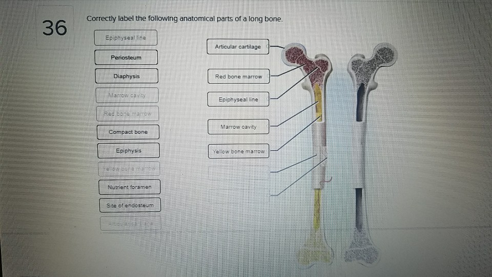
Solved Correctly Label The Following Anatomical Parts Of Chegg Com from d2vlcm61l7u1fs.cloudfront.net The delicate connective tissue layer lining the inside surface of compact bone. The end of the long bone is the epiphysis and the shaft is the diaphysis. When osteoclasts start removing less bone, or osteoblasts start adding more bone, the. Long, short, flat, irregular and sesamoid. Furthermore, on histological sections, fluorescently labeled lin−sca1+kit+ hspc from. Review of long bone anatomy: Long bones, especially the femur and tibia, are subjected to most of the load during daily activities and they are crucial for skeletal mobility. Observe regions of trabecular bone and cortical bone in this specimen.
See bone and cartilage development.
Label the structures of a long bone medullary epiphyseal cavity line spongy articular bone cartilage periosteum compact bone endosteum. (a) growing long bone showing epiphyses, epiphyseal plates, metaphysis and diaphysis. The periosteum is the membrane surrounding the exterior surface of all bones, except the. Labeling portions of a long bone learn with flashcards, games and more — for free. The diaphyseal bone marrow of long bones in these rats sequentially showed three different processes of chronic pathological changes, which, however, partly overlapped each other. Bone and cartilage at rosalind franklin university these pictures of this page are about:long bone endosteum. Long bone endosteum (page 1). Review of long bone anatomy: See bone and cartilage development. The delicate connective tissue layer lining the inside surface of compact bone. Osteoclasts on the inside in the endosteum remove this bone to maintain the bone diameter. This endosteal surface is usually resorbed during long periods of malnutrition, resulting in less cortical thickness. They are one of five types of bones:
Among these cells, you can find the bone stem cells, the ones that are going to further develop into osteoblasts and osteoclasts long bone labeled. (b) mature long bone showing epiphyseal bone tissue that is found in the periosteum, endosteum, suture, and periodontal membrane (ligaments) is an example of intramembranous bone.


0 Komentar